… reaching into the intestinal lumen? They belong to dendritic cells and are depicted also in a nice review about immune responses to commensal and environmental microbes
…. colonization of germ-free mice with B. fragilis expressing PSA results in increases in the number of CD4+ T cells and skews the CD4+ T cell phenotype toward the production of T helper type 1 (TH1) cytokines … On the basis of those results and the observation that antibiotic use may correlate with development of allergic diseases, it was postulated that the presence of diverse commensal populations may thus enhance TH1 and diminish TH2 CD4+ T cell differentiation
which is an interesting explanation that goes back to 3 references – one about the love-hate relationship between LPS and the immune system and two papers by Noverr – it seems that I missed these papers altough they are important to understand our previous findings.
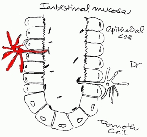
My question, however, was: Can you see this neat little finger of the DCs? I am citing here another review – box 1
DCs actually extend their processes through the tight junctions of epithelia … DC capture of antigens from the environment even when there is no overt infection or inflammation, probably allowing for the silencing of the immune system to harmless environmental antigens. DCs at body surfaces can function locally, for example, to convert vitamins A and D to active retinoic acid and 1,25-dihydroxy-vitamin D3. One consequence of the overlooked metabolic capacities of DCs is to increase the homing of immune cells to that mucosal surface and, in the case of retinoic acid, to help DCs differentiate suppressor T cells, which block autoimmune and inflammatory conditions.
So we have now everything together in the intestinal crypt: bacteria, allergen, D3 prohormone and a cell that senses and metabolizes all of that.