Tag Archives: Vitamin-+-Sunshine

Another path from vitamin D to allergy

As far as I know vitamin D3 influences only RUNX2 expression (RUNX2 has a VDRE, possibly also RUNX3 but not RUNX1?). The RUNX factors

colocalized in common subnuclear foci. Furthermore, RUNX subnuclear foci contain the co-regulatory protein CBFβ, which heterodimerizes with RUNX factors, and nascent transcripts as shown by BrUTP incorporation. These results suggest that RUNX subnuclear foci may represent sites of transcription containing multi-subunit transcription factor complexes.

Variants in RUNX1 have already been reported earlier to be weakly associated with IgE serum levels in Korea.

A Nature paper todays reports that Foxp3 controls Treg function by interacting with AML1/RUNX1 – is there any connecting path?

Higher metabolic rate or better signalling

In a forthcoming paper in Allergy (scienceblog:doi:10.1111/j.1398-9995.2007.01437.x:) we will show an association of vitamin D (25-OH-D3) serum levels and allergic rhinitis (AR) mainly in white Caucasians. Here is a supplemental figure that shows the seasonal variation in AR+ and AR- individuals.

Except of the singular peak in white children, I can´t see so much difference – so probably the vitamin D signalling pathway is different in AR.

Figure: Month of examination in allergic rhinitis patients of NHANES III by age age and ethnic background – no clear effects by higher metabolism.

metaborsignal.png

Lymphopoetin and allergy

This is to convince me that the thymic stromal lymphopoietin (TSLP) is a master switch in allergy

15 Aug 2004

Skin-specific overexpression of TSLP resulted in an AD-like phenotype, with the development of eczematous lesions containing inflammatory dermal cellular infiltrates, a dramatic increase in Th2 CD4+ T cells expressing cutaneous homing receptors, and elevated serum levels of IgE.

5 Jun 2006

Topical application of the physiologically active ligand [1{alpha},25-(OH)2D3; calcitriol] of the vitamin D receptor, or of its low-calcemic analog MC903 (calcipotriol; Dovonex), induces TSLP expression in epidermal keratinocytes, which results in an atopic dermatitis-like syndrome.

22 Jan 2007

TSLP, synergistically with interleukin 1 and tumor necrosis factor, stimulates the production of high levels of Th2 cytokines by human mast cells (MCs) … TSLP is released by primary epithelial cells in response to certain microbial products, physical injury, or inflammatory cytokines.

23 Jan 2007

Proinflammatory TNFalpha or IL-1alpha and Th2 (IL-4 or IL-13) cytokines synergized to induce the production of TSLP in human skin explants … Our data provide the first evidence of TSLP induction and subsequent DC activation in human skin.

25 Feb 2007

Mice with an IEC[intestinal epithelial cell]-specific deletion of IKKbeta show a reduced expression of the epithelial-cell-restricted cytokine thymic stromal lymphopoietin in the intestine and, after infection with the gut-dwelling parasite Trichuris, fail to develop a pathogen-specific CD4+ T helper type 2 (TH2) response and are unable to eradicate infection. Further, these animals show exacerbated production of dendritic-cell-derived interleukin-12/23p40.

June 2007

… (TSLP) and interleukin-7 share a common receptor chain, IL-7Rα … The gene encoding the IL-7Rα chain is polymorphic, and investigation of inhalation allergic patients compared with controls showed significant association with two alleles at position +1237 and +2087.

Aug 2007

… TSLP represents a master switch of allergic inflammation at the epithelial cell and dendritic cell interface.

Dual vitamin D effects on type 1 diabetes and allergy

Early vitamin D supplementation may have dual effects, protecting against type 1 diabetes and inducing allergy. Is that possible? Yes, individual genes variants may determine the outcome.

In addition to HLA the most prominent type 1 diabetes genes are INS – CTLA4 – PTPN22 – CD25 – IF1H1, the most prominent asthma/allergy genes ADAM33 – DPP10 – IL12 – IL4 – GPR154 – FLG. I can´t see any joint master regulator, however, we know that

Could that be a model to explain the dual involvement of vitamin D in both diseases?

Asthma – a disease of the gut

When starting in the asthma field in 1989, the textbooks told me that asthma is a disease of the lung. Some years later, asthma turned out to be a disease of the bone marrow cells. More recently, I raised the question if asthma could be even a disease of the gut – our largest immunological organ being frequently exposed to allergen & plenty of immunological active substances. Although on the different track (vitamin A) also other authors now think of an early impaired immune gut response.
Rather unexpected for me was a study in J Steroid Biochem Mol Biol that 1,25(OH)2D3 inhibits in vitro and in vivo intracellular growth of the parasite Toxoplasma gondii. yea, yea.

Genes wanted

The NIH and Jackson ask for nominations of their gene targeting approach (see also A mouse for All Reasons and my previous comment on the 3 R)

KOMP is a trans-NIH initiative to generate a public resource of mouse embryonic stem (ES) cells containing a null mutation in every gene in the mouse genome. Both conditional and null knockouts are being generated. The purpose of this form is to gather input from the scientific community on which genes should have the highest priority for being knocked out.

The Cell paper also explains the hard to understand differences in knockouts

  1. targeted deletion
  2. targeted conditional
  3. trapped conditional

although I still have semantic problems to understand the nomenclature. Anyway my whishlist – you can do me a favor by voting for CYP27B1, VDR, CYP24A1, OPN, IL4, IL5, IL10, IL12, IL13, FLG, CCR5 and CCR9.

Addendum

You can also leave some input at the Environmental Genome Project.

Let vitamin D shine in

said the Denver Post giving a nice overview of vitamin D research. This was just 1 day too early for a fascinating nature immunology paper (scienceblog:doi:10.1038/ni1433:) that links for the first time natural sunlight induced vitamin D action on dendritic cells. Seems that D3 will influence homing of T cells – we are again at “Licht und Leben“, yea, yea.

Addendum

Vitamin D and asthma

My previous work on vitamin D focused mainly on allergy but according to new research of Bosse et al vitamin D also stimulates bronchial smooth muscle and airway remodeling

Genetic variants in the vitamin D receptor (VDR) gene were recently associated with asthma. The biological mechanisms explaining this association is unknown, but are likely to involve many cell types given the pleiotropic effect of its ligand … The most significant network of up-regulated genes included genes involved in morphogenesis, cell growth and survival as well as genes encoding structural proteins, which are potentially involved in airway remodelling.

Another study published more or less at the same time by Wittke shows

Conversely, WT splenocytes, Th2 cells and hematopoetic cells induced some symptoms of experimental asthma when transferred to VDR KO mice, but the severity was less than that seen in the WT controls … Lipopolysaccharide (LPS) induced inflammation in the lungs of VDR KO mice was also less than in WT mice. Together the data suggest that vitamin D and the VDR are important regulators of inflammation in the lung…

Unripe

A key event in the vitamin D hypothesis of allergy induction is the immature state of dendritic cells. So far, maturity has been mainly described in terms of reduced expression of cell surface marker like CD80. A new study in Nature now further unravels how the capacity of DC to present antigen may be disturbed. Ubiquitination – the covalent attachment of ubiquitin polymers – of the MHC II ß chain ceases on maturation allowing the transport from endosomal compartments to the plasma membrane. Immature cells seem to be capable to some level of peptide-MHC interaction (at least for some selected antigens) although this process is greatly enhanced by maturation of DCs. Semi-maturity is believed to be an important inetrim stage where at least an earlier review argued

we propose a model in which steady-state migration and partial maturation (semi-maturation) of DCs is embedded as a major component within immune homeostasis, established for permanent and active tolerance induction against self-antigens derived from peripheral tissues by inducing antigen-specific CD4+ Tr cells. Semi-maturation induced by proinflammatory cytokines, such as TNF-alpha, seems to represent a unique developmental tolerogenic stage for DCs, which is based on the absence of proinflammatory cytokine production, despite high expression of MHC II and costimulatory molecules.

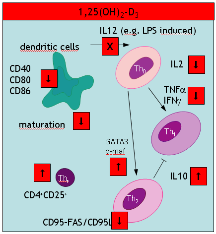
Another interesting study in the J Immunol – coined “alternatively activated dendritic cells” the authors probably talk about the same immature cells (compare with my cartoon summarizing a 2002 paper in Trend Mol Med). These immature DCs secrete high levels of IL10 (a paradox discussed in my most recent paper). In addition these DCs produce low amounts of IL12p70, TLR4 and CCR7. What was new to me, was an impressive list of pharmacological agents that suppress DC development: aspirin (also paracetamol?), corticosteroids, cyclosporine A, rapamycin (also other antibiotics?), and finally mycophenolate mofetil.

There is also an update of the IL10 paradox: Allergic sensitization may be down regulated by CD40 AGONISTs independent of IL10!

Finally, I would like to understand what immature really do after encountering allergen exposure. A new paper in nature immunology says that

immature DC are also thought to carry antigen to lymph nodes and to interact with naive T cells but without a previous maturation stimulus, those interactions result in abortive activation of the T cells, which can be eliminated, rendered unresponsive or induced to differentiate into regulatory T cells.

which still does not answer my question.

Vitamin D actions
vd_immun.png

Yea, yea.

Beauty is skin deep but ugly goes all the way to the bones

Sorry, the original title of this paper is “Nothing but skin and bone“. It is an excellent JCI review of two diverse systems that do not seem to have so many common denominators. As the authors state a recurring theme is Wnt (a family of secreted glycoproteins) signalling during early patterning and for morphogenesis of skin appendages – common mesenchymal progenitors support both tissues. What the authors neglected is the effect of vitamin D signal in skin and bone, yea, yea.

Resurrection or reanimation of old myth?

JCI has a paper about resurrection of vitamin D deficiency (more about the author at 1, 2 and 3). The author uses references 46-59 to underpin his opinion that rickets has again become an epidemic. These references are from 1992, 1984, 2005, 2000, 1987, 1987, 1989, 1994, 1989, 2001, 1998, 2001, 1987, 2006. So nothing really new – no prospective study, no systematic survey, just a few isolated reports. Sure, that there might be a rickets problem in a selected areas or in minority groups, but there is no world-wide epidemic. He argues also that many (if not all) studies show “low” serum 25-OH-D3 values. Is this chasing a phantom? A more systematic study concludes that rickets in Africa is more a disease of calcium deficiency. The JCI article is particular poorly written when it comes to immunological effects; asthma is misquoted from the Camargo study (which is subject of my review at Pediatric Allergy. Nay, nay.

Nothing but skin and bone…

… was the title of a recent editorial in JCI. Clearly that’s not true for vitamin D as you may know of the many immunological functions. A new study now shows, that mice get atopic dermatitis by applying vitamin D ointment. A EJCN paper published on the same day concludes that “the national fortification of fluid milks and margarines with vitamin D safely improved the vitamin D status of children”. Nay, nay.