Tag Archives: allergy

Warum es in der DDR so viel weniger Allergien gab

Ich hatte direkt nach der Wende in Halle und Leipzig die erste Ost-West Allergiestudie* durchgeführt und dabei die verblüffende Entdeckung gemacht, dass es im Osten nicht mal halb so häufig Allergien gab.


https://www.atsjournals.org/doi/pdf/10.1164/ajrccm.149.2.8306030


Der Grund dafür war uns zunächst nicht klar, aber dann stelle sich doch bald heraus, daß es wohl die unterschiedliche Vitamin D Prophylaxe war – im Westen gab es tägliche niedrige Dosen ab der ersten Lebenswoche, im Osten wurde ab dem 2. Monat wenig   hohe hohe Einzeldosen verabreicht. Wenn Kinder im Osten  krank waren, dann fiel auch immer wieder die eine oder andere Dosis aus, auch wurde das Schema nach meiner Recherche nicht immer komplett durchgezogen. Leider haben wir uns damals aber nur die Impfpässe angesehen, nicht aber die im Nachinein wichtigeren Wiegekarten.


DDR Wiegekarte. Quelle https://www.ddr-museum.de/de/objects/1011262
Westdeutsches Untersuchungsheft U2. Quelle https://www.g-ba.de/downloads/83-691-452/2017-05-11_GBA_Kinderuntersuchungsheft_Web-WZ.pdf


Die Befürchtung der Professoren Mai und Beuren in dem alten SPIEGEL Artikel über Vitamin D Nebenwirkungen haben sich zum Glück nicht bewahrheitet. Dafür aber stellte sich dann aber eine  unerwünschte immunologische Wirkung heraus die damals noch nicht bekannt war.

Mit dem aus England bzw Amerika importierten Schema zur Supplementierung  stiegen die Allergien an. In der BRD war das ab den frühen 60er Jahren , wie überhaupt die englischsprachigen Länder immer die höchsten Allergiehäufigkeiten hatten, da sie wegen der häufigen Rachitis auch viel konsequenter supplementierten (die Rachitis hiess früher einmal auch “englische Krankheit”).

Mit dem Mauerfall 1989 setzte der Anstieg dann auch in der ex DDR ein und erreichte nach 10, 20 Jahre  das Westniveau . Leider wurden unsere Warnungen vor einer zu frühen Supplementierung nicht ernst genommen, der Osten Deutschlands hat – wie so vieles andere auch – das Schema aus dem Westen übernommen und den Preis dafür mit ebenfalls hohen Allergieraten bezahlt.

Der Mechanismus der Allergieentstehung ist dabei nur teilweise aufgeklärt: Vitamin D ist jedenfalls immunsuppressiv mit vielfacher Auswirkung auf B und T Zellen was seit  dem Nachweis des Vitamin D Rezeptors auf diesen Immunzellen wissen. Die Supplementierung stört offensichtlich die initiale Klassifikation ob ein Protein harmlos oder allergen ist.

Warum es aber auch schon 1989 Allergien im Osten gab? Nun, es war ja keine Vitamin D freie Zone, offensichtlich sensibilisiert man sich auch noch ausserhalb des überkritischen Intervals in den ersten Lebenswochen.

Allergy GWAS hits in VDR binding sites

It seems that I missed an interesting 2017 paper that looked for disease-associated SNPs in canonical DR3 motifs. Only 7 out of 211 traits showed significant hits, one of these was self-reported allergy. When annotating these SNPs, there are only two genes: LINC00299 and TLR1

hg38 position
rs10174949 2:8302018 LINC00299
rs10178845 2:8303773 LINC00299
rs5743566 4:38804221 TLR1
rs2101521 4:38809830 TLR1
rs5743565 4:38804262 TLR1
rs45588337 4:38805607 TLR1
rs55830619 4:38805643 TLR1

So are TLR1 & LINC00299 variant carriers more susceptible to vitamin D induced allergy?

LINC00299 (Long Intergenic Non-Protein Coding RNA 299) is a RNA Gene of largely unknown function, associated so far with allergy only on a genetic level in Framingham,  href=”https://pubmed.ncbi.nlm.nih.gov/23817569/”>23andme and other studies. We don’t know so much here, the function of the long non coding RNAs

depends on subcellular localization. Depending on their niche, they specifically interact with DNA, RNA, and proteins and modify chromatin function, regulate transcription at various stages, forms nuclear condensation bodies and nucleolar organization. lncRNAs may also change the stability and translation of cytoplasmic mRNAs and hamper signaling pathways. Thus, lncRNAs affect the physio-pathological states and lead to the development of various disorders, immune responses, and cancer.

The TLR1 genetic association is found by many genetic studies, while the clinical association is probably more by an infectious origin. TLR1 is a pattern recognition receptor with a specificity for gram-positive bacteria and also included in my forthcoming exome paper as a protective factor for asthma/allergy.  And we are also close to  my earlier review of vitamin D, the microbiome and allergy…

Does any co-infection response during first vitamin D exposure influence allergic sensitisation? There are indeed some hints of an short-lived effect of lung group 2 innate lymphoid cells (ILC2s)

Laboratory mice cohoused for 2 weeks had impaired ILC2 responses and reduced lung eosinophilia to intranasal allergens, whereas these responses were restored in mice cohoused for ≥2 months. … These findings suggest that ILC2s respond dynamically to environmental cues and that microbial exposures do not control long-term desensitization of innate type 2 responses to allergens.


Hygiene hypothesis hyperbole

Having written about the hygiene hypothesis, I missed a PNAS News feature even some years ago.

Again: The hygiene hypothesis was not born by David Strachan and well, there are more cracks of the hygiene hypothesis.

“The trouble is, as soon as you use the words ‘hygiene hypothesis,’ the word hygiene prejudges what the cause is,” says Bloomfield. To the public, “hygiene” is interpreted as personal cleanliness: washing hands, keeping food clean and fresh, sanitizing the home. However, because the hypothesis has been largely uncoupled from infections, the idea that we need to be less hygienic is wrong. Relaxing hygiene standards would not reverse the trend but only serve to increase the risks of infectious disease, says Bloomfield. The term “hygiene hypothesis” also fails to incorporate all of the other factors now linked to the increase in immunoregulatory diseases.

I expect that five years after mandating “super hygiene” during COVID-19 we can finally bury the hygiene hypothesis.

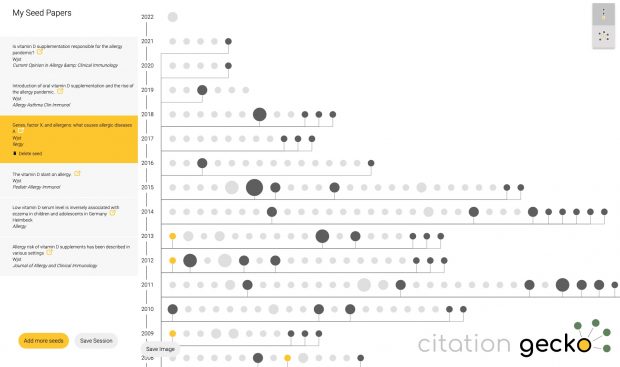
Not too bad: Citation Gecko

Just tried citationgecko.com on a topic that I have been working on for 2 decades. It will find rather quickly the source  paper, much faster than reading through all of it. Unfortunately reviews are rated to be more influential than original data as Citation Gecko picks articles with many references.

Parental allergy history at farms

A recent paper on the bias of farming studies did not discuss a  healthy worker although this is being a reasonable explanation.

So let’s have a more detailed look here at farm parents. It is an important question if parents are also “protected” (or if some affected parents just moved away).

AFAIK there are 7 studies dealing with this question. Continue reading Parental allergy history at farms

What is needed is a confrontational approach

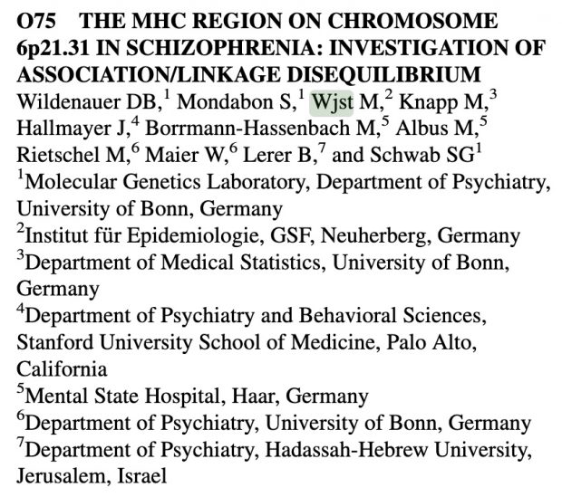
Google showed me today a long forgotten abstract. Although I am not an expert in schizophrenia we collaborated two decades ago on this particular chromosomal region.

What is even more remarkable is the challenging keynote of the Xth World Congress of Psychiatric Genetics in The Palais des Congre ́s Brussels, Belgium October 9 –13, 2002 by Irving Gottesman [+2016], the father of epigenetics in psychiatry. He wrote there

We cannot escape the history of our field and are constantly guided today by the accumulation of facts with either positive or negative valences from our past. But when did the clock start—with the domestication of animals, with Galton’s musings and amoral passion for data collection about individual differences in behavior, or with the initially objective scientizing of Mendelism applied to schizophrenia but ending with a Nazi-tainted albatross around the neck of psychiatric genetics. In regard to the long quest for the distal and genetic (partial) causes of mental diseases, the conclusion that both genetic and environmental factors, none yet known in detail, provide the distal causes of mental disorders—that statement is too general to be of use to making further progress. What is needed is a confrontational approach based on evidence collected from competing ‘schools of thought’, and then reconciliation before some kind of omniscient and impartial Science Court.

I couldn’t agree more. What is needed is a confrontational approach based on evidence collected from competing ‘schools of thought’, and then reconciliation before some kind of omniscient and impartial Science Court.

How atopic dermatitis is linked to vitamin D and how IL33 splice variants associate to eosinophil numbers

(first published 12 Dec2020 and revised 10Dec2021)

We had a major discussion right before our 2010 paper where I argued that rare variants should have been included into our asthma/allergy/dermatitis GWAS. Ten years after there is now a nice paper using massive exome sequencing that finally includes them.

It seems that the respiratory tract isn’t so much influenced by rare gene variants but that there is a strong effect in  the immune system.

And there is another interesting fact.

…Surveying the contribution of rare variants to the genetic architecture of human disease through exome sequencing of 177,882 UK Biobank participants …if we look at the …. European population who are carriers of a filaggrin (FLG) PTV, we find those carriers have significantly higher risk for well-known associations, such as dermatitis  … and asthma … Concomitant increases in vitamin D levels suggest … increased sensitivity to ultraviolet B radiation.

So far, I have only assumed an asthma/allergy priming effect of oral vitamin D in the newborn gut. This paper now argues for an increased vitamin D sensitivity also in the skin of  FLG dermatitis patients which is interesting given the largely contradictory data of serum vitamin D and atopic dermatitis. Maybe dermatologists should focus their research more on skin and local vitamin D turnover?


-II-


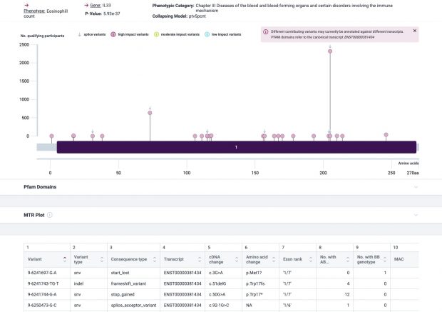
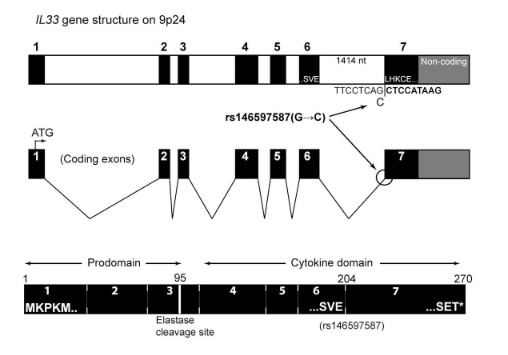
The most prominent IL33 variant carried by over 2,300 people is splice acceptor 9-6250473-G-C followed by 600+ individuals with splice donor 9-6250600-G-T.

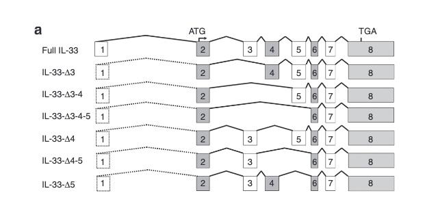
There are not too many carriers of this variant by the sheer amount of 177,882 participants. We nevertheless know already something about the seven IL33 splice variants since 2012.

Novel Splice Variants of IL-33: Differential Expression in Normal and Transformed Cells Journal of Investigative Dermatology (2012) 132, 2661–2664; doi:10.1038/jid.2012.180

with updates  in  2016

Gordon, Alternative splicing of interleukin-33 and type 2 inflammation in asthma, PNAS 2016

as well as in 2017

Fig 3A Smith et al. A rare IL33 loss-of-function mutation reduces blood eosinophil counts and protects from asthma, PLoS Genetics 2017 – describes the splice site as NM_001199640:exon7:c.487-1G>C or rs146597587-C


So I did a sequence match to compare the new finding with these older publications.

own sequence match exon7 using data from dbSNP, UCSC GoldenPath and Uniprot – reference is hg19


Indeed, the 2017 paper already described rs146597587 which is probably identical to the splice acceptor 9-6250473-G-C in Astra UK Phewas (genome positions do not match – I used hg19 while I don’t know the Astra reference) . Astra says also c.613-1G>C while rs146597587 is just upfront of my codon 205 (3*205=615) whatever that means.

The Astra UK Phewas at least confirms the Iceland paper above

rs146597587-C associates with lower eosinophil counts (ß= -0.21 SD, P = 2.5×10⁻¹⁶, N = 103,104), and reduced risk of asthma in Europeans (OR = 0.47; 95%CI: 0.32, 0.70, P = 1.8×10⁻⁴, N cases = 6,465, N controls = 302,977). Heterozygotes have about 40% lower total IL33 mRNA expression than non-carriers and allele-specific analysis based on RNA sequencing and phased genotypes shows that only 20% of the total expression is from the mutated chromosome. In half of those transcripts the mutation causes retention of the last intron, predicted to result in a premature stop codon that leads to truncation of 66 amino acids.

So it is basically a rediscovery meaning that we reached saturation.

Vitamin D traces in later life

This is basically an update of my 2017 Allergy paper where I asked about sequelae of early vitamin D supplementation.

Two extensively examined hypotheses are the hygiene hypothesis (lack of protective bacterial exposure which leads to subsequent allergy) and the vitamin D hypothesis (early vitamin D supplementation sensitizes newborns against allergens) …  The interesting question is: Are these concepts exclusive? …  There is some preliminary evidence that – like many other environmental factors –vitamin D may modify the human microbiome.

Only yesterday a paper popped up during a presentation of Amelie Baud about the influence of social partners and the gut microbiome. This 2018 study  tested gut microbial composition from 16S rRNA sequencing during the first year of life and subsequent risk of asthma in 690 participants

1-year-old children with an immature microbial composition have an increased risk of asthma at age 5 year … the microbial composition was not affected by maternal asthma status suggests that only susceptible children, exposed to inappropriate microbial stimulation during the first year of life, may express their inherited asthma risk …. The five most discriminating indicator OTUs for each cluster were identified for PAM cluster 1 as Enterobacteriaceae, Staphylococcus, Streptococcus, Bifidobacterium and Enterococcus, and for PAM cluster 2 as Faecalibacterium, Bacteroides(x3), and Anaerostipes … the risk of developing persistent asthma was increased (adjusted hazard ratio (aHR) 2.87 (1.25−6.55), P = 0.013) if the microbiome remained in PAM cluster 1 at age 1.

IMHO this doesn’t look very much like direct microbiome effects but some colliding  factor. The authors discuss cesarean section-birth and antibiotics as relevant factors while I wonder why the last author (who is a known pro vitamin D lobbyist ) doesn’t take into account vitamin D here?

My 2017 review summarized only early results, where there are now many more robust studies like the 2019 Naderpoor study

there was a significant association between community composition and vitamin D supplementation at the genus level. The vitamin D group had a higher abundance of genus Lachnospira, and lower abundance of genus Blautia (linear discriminate analysis >3.0). Moreover, individuals with 25(OH)D >75 nmol/L had a higher abundance of genus Coprococcus and lower abundance of genus Ruminococcus compared to those with 25(OH)D <50 nmol/L.

or the 2020 Singh paper

Vitamin D supplementation significantly increased gut microbial diversity. Specifically, the Bacteroidetes to Firmicutes ratio increased, along with the abundance of the health-promoting probiotic taxa Akkermansia and Bifidobacterium. Significant variations in the two-dominant genera, Bacteroides and Prevotella, indicated a variation in enterotypes following supplementation.

So is the microbiome just an indicator of vitamin D exposure in genetic susceptible children?

When a scientific journal is modifying your images

This content has restricted access, please type the password below and get access.


Blaming genes for resistance to environmental stressors

This something that I always avoided in human research – blaming genes for resistance to environmental stressors.

Nevertheless a Californian group (https://doi.org/10.1371/journal.pgen.1008528) now tested 101 mouse strains for lung resistance with exposure to diesel exhaust particles (DEP). After sensitizing the animals with dust mite and aluminium they could also test metacholine hyperreactivity (AHR).

Strains that exhibited the highest lung resistance after control exposure were not necessarily the same as those with high lung resistance after DEP exposure. It is unclear which strain was used for the consecutive GWAS. Did they put all mice into one cage for that?

The metacholine AHR GWAS results are not very impressive. And there seem to be also errors, as for example the lead SNP on chr 19 (rs51547574, near IL33) is shown with different allele frequencies in text and Fig 2.  As the expression quantitative trait locus (eQTL) for Il33 is not in the lung, I think there is nothing to memorize here – IL33 is just a gatekeeper for surface integrity.

In a next step I wouldhave expected a GWAS for resistance change after DEP but FIG 3 only gives the result of Δ AHRDEP—AHRPBS data at an abitrary methacholine dose of 10mg/ml. The identifed locus could be interesting but as the LD there is rather high without any corresponding eQTL (I always wondered why there has never been a significance threshold for LD blocks, only for isolated SNPs?), the logic of the paper is broken here. Induction of lung resistance by DEP was significantly blunted in Dapp1-/- female mice? What about male Gm5105-/- Mttp-/- , and Lamtor3-/- animals?

Hopefully nobody else will now try to find diesel, ozone, NOx resistance genes in humans as this is not a a scientific but a political issue…

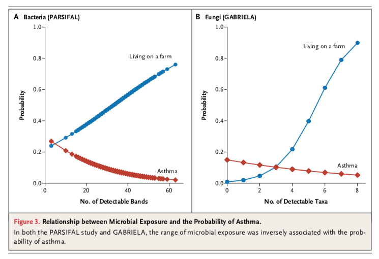
What is wrong with the 2011 NEJM paper?

N Engl J Med 2011;364:701-9 is another paper with 1000+ citations  that had a lasting impression on some but not all people.

First, I can’t remember of any study with such an enormous selection bias  where >94% of individuals have been lost.

Second, we should not forget that farm is not protective per se – farmers may just avoid a known allergy risk factor.  PARSIFAL participants in this study included Steiner schools — anthroposophic medicine mostly avoids vitamin D (ref). This is of course a major issue for any cross-sectional study that doesn’t take into account the temporality of events.

Third, in PARSIFAL dust from children’s mattresses were collected by vacuuming — it is not very likely that many helminthic eggs were transported  from stable to bedroom. In GABRIELA, only airborne dust samples  were collected which again may miss helminth eggs although being certainly present in stable dust.

Fourth, more  microbial exposure and more fungal taxa on farms are a trivial finding.

The inverse associations of the diversity scores with asthma were not confounded by status with respect to living on a farm because adjustment did not change the respective point estimates for asthma (Table 2), although the associations became nonsignificant.

Small sample size, borderline p-values even after a long fishing expedition?

What do these strange “probability” plots  really show – the probability of asthma or the probability to live on a farm?

N Engl J Med 2011;364:701-9 Figure 3 Does it refute any general effect of diversity?

The plots are misleading if adjustment for farm living does not change the parameter estimates for bacterial/fungal diversity.

Sixth – even many years later, the main findings of this study have not been independently replicated. There is not any single study that shows listeriosis (Listeria) or diphtheria (Corynebacterium)  to be protective.

medRxiv

ArXiv is operational since 1991, bioRxiv since 2013 and since 2019 there is now also medrxiv. More details  at https://www.bmj.com/content/365/bmj.l2301

The main arguments in favour of sharing work in its preliminary form are, firstly, that science works faster if work is made available sooner after it is completed and, secondly, that articles are improved by feedback from a wider group of readers, alongside formal peer review by a few experts. Simple estimates suggest that halving the delay to sharing a research result can double the speed at which research progresses. Ambitious research funders are now embracing preprints and other measures that aim to accelerate the pace of research.

Although there was a mixed reception in the beginning, see Science back in 2017

MedArXiv will have a hard time attracting preprints if mainstream medical journal editors decide they won’t publish final versions of the papers. Currently, The BMJ and The Lancet are among the few medical journals that have explicitly said that posting a preprint doesn’t preclude publication; Nature and Science, which both occasionally publish medical studies, have the same policy. But at the JAMA Network, which publishes a dozen journals, the issue is hotly debated.

@medRxiv opened on June 6. So far they have only 304 followers on Twitter (and no allergy paper in the archive).

As the current “Allergy” editor and the publisher (John Wiley and Sons A/S)  agreed to preprints last week, I have submited now my first preprint paper. Therefore, there are now 305 followers and 1 allergy paper :-)

IL33, allergy and helminths: Shot in the leg?

Ever since our NEJM paper in 2010 that showed an IL33/ST2 association there are new studies on IL33.

Grotenboer 2013 did a functional annotation of the gene and it’s receptor in humans while there is no more doubt about the involvement of IL33 in human allergy.  Right now IL33 suppression is already used as an experimental screening test for allergic reactivity with ongoing phase II studies of anti-IL33 or anti ST2. Good IL33 reviews can be found for example in frontiers in immunology by Tataori et al. or in nature immunology by Smith.

These reviews do not tell you so much about the regulation while regulation has recently elucidated by Gour et al. who describe a tropomyosin–dectin-1 interaction of the human host. Why is tropomyosin such a frequent target of human IgE?

Muscle protein tropomyosin is an important IgE target in a number of nematode infections; Onchocerca volvulus ; Ascaris lumbricoides; Anisakis simplex; and tropomyosin from the blood fluke Schistosoma mansoni is also a human IgE antigen. Tropomyosin is highly conserved across many invertebrates and is responsible for much of the IgE cross-reactivity between Ascaris and dust-mites.

I haven’t found any good  answer to this question. As tropomyosin affects contractility – this seems like “shooting into the leg” of worms whenever they attempt to invade.

Maybe Gour et al. did not know the earlier dissertation from Berlin that already showed a reduced inflammation in the OVA mouse model by administration of recombinant tropomyosin.

The broad cross reactivity to tropomyosin gives rise to the question if helminth tropomyosin could induce allergic reactions to itself and/or tropomyosin of different organisms. Considering the fact that filarial nematodes express tropomyosin on their surface […] and that the continuing turnover of microfilariae confronts the host with relevant amounts of tropomyosin makes this question even more appropriate.

Worms seems to be attacked by anti-worm-surface-tropomyosin IgE whenever the worm tries to invade  the epithelium during an acute infection. During invasion extracellular IL33 is cleaved into a shorter form with enhanced activity attracting more immune cells.
During chronic infestation nothing happens as long as the worm does not invade and doesn’t trigger any IL33 alarmin. As there is continuous tropomyosin antigen antigen contact, the host is slowly desensitzed, clearing IgE in favor of IgG4.

Is this also a model that explains allergy? We don’t know the details but maybe this antigen recognition / response system is being disturbed where allergens like Der p1 mimicking a worm infection by tropomyosin can trigger the allergic reaction in particular as Der p1 a cysteine protease also mimicks an invasion signal.

23.12.2019 Addendum

Parasite tropomyosin ist detected in in 55%-62% of patients (cockroach tropomyosin rPer a 7, Ascaris tropomyosin rAsc l 3).

People who behave fraudently

Richard Smith 3:58 “People who behave fraudently tend to behave fraudently in all aspects of their live”

8:05 “Universities have no interest to discover fraud” 10:42 “Journals don’t have the legal standing” 10:58 “we need some kind of national bodies”

A step forward in allergy research

The most recent paper of my Australian collaborators is a relevant step forward: Polymorphisms affecting vitamin D-binding protein modify the relationship between serum vitamin D (25[OH]D3) and food allergy. Basically they show an

… association between serum 25-hydroxyvitamin D3 (25[OH]D3) levels and food allergy at age 1 year (338 challenge-proven food-allergic and 269 control participants) and age 2 years (55 participants with persistent and 50 participants with resolved food allergy)… Analyses were stratified by genotype at rs7041 as a proxy marker of DBP levels… Low serum 25(OH)D3 level (<50 nM/L) at age 1 years was associated with food allergy, particularly among infants with the GG genotype (odds ratio [OR], 6.0; 95% CI, 0.9-38.9) … Maternal antenatal vitamin D supplementation was associated with less food allergy, particularly in infants with the GT/TT genotype (OR, 0.10; 95% CI, 0.03-0.41)… This increases the biological plausibility of a role for vitamin D in the development of food allergy.

Maybe it would be helpful to have also “real” DBP levels for estimating bioavailability (and even data of supplement use) but already the reported results are another strong argument for the vitamin D – allergy axis. This is also largely in line with what I predicted back in 2012

Both vitamin D insufficiency and vitamin D supplementation have been linked to allergy and asthma. This apparent paradox is explained by epigenetic programming in pregnancy by low vitamin D levels and the excessive high supplementation in the newborn period.

Maybe I should have emphasized that genetic variants in the vitamin D pathway are also important for biological effects.