Category Archives: Vitamins

Vitamin D responder: Check CD14 methylation

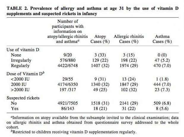
A clinical study in 2015 already showed, how to recognize vitamin D responders using a preselected gene set based on VDR chip seq data.

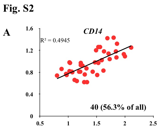
Vitamin D3 is a pleiotropic signaling molecule that has via activation of the transcription factor vitamin D receptor (VDR) a direct effect on the expression of more than 100 genes. The aim of this study was to find transcriptomic and clinical biomarkers that are most suited to identify vitamin D3 responders within 71 pre-diabetic subjects during a 5-month intervention study (VitDmet). In hematopoietic cells, the genes ASAP2, CAMP, CD14, CD97, DUSP10, G0S2, IL8, LRRC8A, NINJ1, NRIP1, SLC37A2 and THBD are known as primary vitamin D targets […] only 39-44 (55-62%) of the study subjects showed a highly significant response to vitamin D3, i.e., we considered them as “responders” … genes were expressed but in a wide range that differed up to 327-fold between the most prominently (CD14) and the lowest expressed candidate (CAMP)

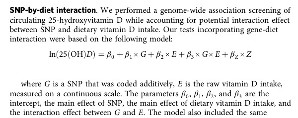
y-axis end/start CD15 serum, x-axis end/start 25-OH-D3 

In new editorial, the same authors argue that the vitamin D response index is an epigenetic property of an individual that may not change at all. Indeed, changes in the epigenome, such as methylation of genomic DNA is an essential prerequisite for initiating gene transcription. The concept of an individual vitamin D response therefore has a lot of merits and is able to resolve a long controversy which serum vitamin D levels are sufficient.

CD14 is a particular interesting gene. As we have learned in allergy research, however, methylation status is not stable over time, it increases slightly over the first decade, possibly as vitamin D sensitivity decreases??

The average increase in CD14 methylation from 2 to 10 yr (n = 153) was 1.3% (from 5.5% to 6.8%, p = 0.001)

This difference isn’t really huge while also the time spent outdoors seem to be relevant. CD 14 methylation may even influence SNP association results

rs2569191, rs5744455, and rs2569190 were associated with sCD14 levels at birth and 2 years, but only rs5744455 was associated with sCD14 levels at 10 years. CD14 methylation increased significantly from age 2 to 10 years.

So CD14 methylation looks like an interesting indicator and may even have biological relevance itself as CD14+ monocytes can differentiate into a host of different cells.

Death of a newborn following vitamin D drops

aerzteblatt.de, thejournal.ie, bbc.com, reuters.com, lemon.fr (with video) report that

France has moved to suspend sales of a vitamin D medication following the death of a baby who suffocated after being given the liquid supplement, health authorities said today. France’s ANSM agency that oversees the safety of medicines and health products said it had taken the measure “as a precaution” after investigations showed “a probable link between the death and the administration of Uvesterol D”.

Indeed vitamin D is used as a rodenticide with high toxicity. The death of the baby was probably not a direct effect of vitamin D but induced by the kind of application. In any case, I would avoid any oral application of an immunosuppressive hormone to a healthy newborn.

Rat race

While working on a review about vitamin D and the microbiome, I came across an interesting article Immune-Microbiota Interactions: Dysbiosis as a Global Health Issue

Recent research, however, demonstrated that a number of specific interventions can lead to (partial) primary prevention of allergy, especially of atopic dermatitis (AD) and food allergy (FA). Three types of primary prevention strategies have been successfully studied: early administration of bacterial products (most studies are on probiotics), early moisturizing in infants at risk for AD and early exposure to allergenic foods (peanut and egg).

I am not so much convinced of any successful probiotics research that prevents all kind of allergy (ref, ref, just to name two). The interesting point, however, is the new recommendation to early exposure of allergenic foods. Does earlier exposure mean less exposure under vitamin D suppression that shouldn’t start before week 6?

Low serum vitamin D – reverse causation or true risk factor?

Even with thousands of studies, one of the most basic questions in vitamin D research is being unanswered. Low serum 25(OH)D3 – is it reverse causation or a true risk factor? As plasma 25(OH)D3 can be easily determined, it became the standard measurement for vitamin D supply. Unfortunately numerous other factors – season, genetics, sex, age, race – all influence serum 25(OH)D3 levels.

Then in 2011 David Reid et al. published a paper of 25(OH)Dfollowing up changes during acute inflammatory response after knee surgery. In essence, plasma concentrations of 25(OH)D decreased after an inflammatory insult and are not a reliable measure of 25(OH)D status in subjects with a significant systemic inflammatory response. This observation is being confirmed in the meantime by 2 further studies (Barker 2012 and Waldron 2013). I think this is a clear result now.

I have never been convinced that Mendelian randomization will help here at all – as done in earlier studies. These are clearly situations where Mendelian randomization does not work as shown  by Smith and Ebrahim back in 2004

—failure to establish reliable genotype (seldom)
—intermediate phenotype genotype—disease associations (frequent)
—confounding of genotype—intermediate phenotype—disease associations (unclear)
—horizontal pleiotropy and multi-functional genes (frequent)
—canalization and developmental compensation (unclear, but expected to be frequent)
—Lack of suitable polymorphisms for studying modifiable exposures of interest (no more a problem)

So, 25(OH)D3 is more an acute phase indicator where low levels in inflammatory diseases is effect not a cause but an effect. A recent review concludes therefore

Reversed causality is described as a possible factor interfering with the correct assessment of the Vit D status. It is concluded that further widespread fortification of foods and stimulation of supplement use should be reconsidered.

It is, however, unclear if low 25(OH)D levels is due to an increased demand (as the vitamin D lobby argues), a shift in free/bioavailable metabolite by hemodilution, binding protein capacity (what I am expecting) or just some unknown further factors.


Vitamin D Vergiftung bei Weidetieren

Auch Kühe können erhöhte Vitamin D Werte haben, wenn sie Goldhafer fressen, der ab 500m im alpinen Raum auftritt.

Trisetum flavescens / Goldhafer ist zwar kein bevorzugtes Futter und wird nur gefressen, wenn das Angebot von anderen Gräsern knapp ist. Der Hafer enthält aber Calcitriol, das aktive Vitamin D Hormon (ansonsten kommt Vitamin D in der Nahrungskette nur als Ergocalciferol in sonnenbestrahlten Steinpilzen, Pfifferling, Spinat, einigen Kohlarten und Hefe vor. Lebensmittel mit hohem Gehalt tierischen Ursprungs sind Hühnereier, fettreiche Fische wie Lachs, Forelle, Thunfisch).

Sensationell ist jedenfalls der  Hormongehalt des Hafers, den es sonst in keinem Lebensmittel gibt. Das bayrische Landesamt für Umwelt 2007 weiss auch, wo besonders viel Goldhafer in Bayern zu finden ist – südlich des Chiemsees

Einziger Fundort [von Trisetum flavescens ssp. purpurascens (Goldhafer)] im Landkreis am Sagberg bei Frasdorf in Goldhaferwiesen bei 820 m, an den rotbraunen, kräftigen Fruchtständen und kräftigeren Blattspreiten gut kenntliche Unterart des Goldhafers, bisher nur bekannt und zahlreich nachgewiesen aus höheren Lagen der Allgäuer Alpen. Außerhalb davon nur noch drei Meldungen aus tieferen Lagen im Rahmen der ABK (außer diesem sonst nur noch Einzelnach- weise aus den Landkreisen Lindau und Garmisch-Partenkirchen).

map

Why allergy and vitamin D trials failed so far

The following figure highlights the exposure scheme in the three published vitamin D / allergy clinical trials so far: Grant 2016, Litonjua 2016 and Chawes 2016.

sc

None of them found anything important, maybe due to the design flaw in all these studies?

All three studies have been designed with the assumption of a beneficial vitamin D effect.

They are dose finding studies that do not allow to prove or disprove any vitamin D effect.

A step forward in allergy research

The most recent paper of my Australian collaborators is a relevant step forward: Polymorphisms affecting vitamin D-binding protein modify the relationship between serum vitamin D (25[OH]D3) and food allergy. Basically they show an

… association between serum 25-hydroxyvitamin D3 (25[OH]D3) levels and food allergy at age 1 year (338 challenge-proven food-allergic and 269 control participants) and age 2 years (55 participants with persistent and 50 participants with resolved food allergy)… Analyses were stratified by genotype at rs7041 as a proxy marker of DBP levels… Low serum 25(OH)D3 level (<50 nM/L) at age 1 years was associated with food allergy, particularly among infants with the GG genotype (odds ratio [OR], 6.0; 95% CI, 0.9-38.9) … Maternal antenatal vitamin D supplementation was associated with less food allergy, particularly in infants with the GT/TT genotype (OR, 0.10; 95% CI, 0.03-0.41)… This increases the biological plausibility of a role for vitamin D in the development of food allergy.

Maybe it would be helpful to have also “real” DBP levels for estimating bioavailability (and even data of supplement use) but already the reported results are another strong argument for the vitamin D – allergy axis. This is also largely in line with what I predicted back in 2012

Both vitamin D insufficiency and vitamin D supplementation have been linked to allergy and asthma. This apparent paradox is explained by epigenetic programming in pregnancy by low vitamin D levels and the excessive high supplementation in the newborn period.

Maybe I should have emphasized that genetic variants in the vitamin D pathway are also important for biological effects.

5% of methylated sites escape reprogramming – a new allergy research direction

New Scientist Health has a short report how parents’ lives could change children’s DNA.

Azim Surani at Cambridge University and colleagues have demonstrated that some genes in the developing fetus escape the cleaning mechanism. Surani’s team analysed methylation patterns in a type of fetal cell that later forms a fetus’s own sperm or eggs. We would expect these cells to have been wiped clean when the fetus’s epigenome was reset at the early embryo stage. “However, about 2 to 5 per cent of methylation across the genome escaped this reprogramming,” says Surani.

The current wave of interest stems from three new papers: “The Transcriptome and DNA Methylome Landscapes of Human Primordial Germ Cells” by Guo demonstrates

The transcriptome of human primordial germ cells from the migrating stage to the gonadal stage reveals that both pluripotency genes and germline-specific genes are simultaneously expressed within the same individual cells. The global erasure of DNA methylation creates a super-hypomethylated germline genome.

So at week 10 after gestation, all analyzed 233  primordial germ cells lost their parental methylation marks except of 6% of the male and 8% of the female genome (which is a bit larger) . Unfortunately I did not find a list of genes there that have their parental methylation status transmitted.

Tang from a British consortium “A Unique Gene Regulatory Network Resets the Human Germline Epigenome for Development” writes

A unique transcriptome drives extensive epigenome resetting in human primordial germ cells for establishment of totipotency. Some loci associated with metabolic and neurological disorders exhibit resistance to reprogramming and are candidates for transgenerational epigenetic inheritance.

Here evolutionarily young and potentially hazardous retroelements, like SVA, remain methylated ( the number of embryos  being examined is not given). Evolutionarily young and potentially hazardous retroelements, like SVA, remain methylated. When testing for resistant loci, they found that H3K9me3 marked escaping ; resistant regions were also enriched for KAP1 (alias TRIM28) binding sites of ESCs. But still no gene list there.

Sofia Gkountela “DNA Demethylation Dynamics in the Human Prenatal Germline” from the US

performed whole-genome bisulfite sequencing (WGBS) and RNA-sequencing (RNA-seq) of human prenatal germline cells from 53 to 137 days of development. We discovered that the transcriptome and methylome of human germ-line is distinct from both human PSCs and the inner cell mass (ICM) of human blastocysts … Gene expression do not correlate with global changes in DNA methylation.

In this paper finally there is the gene list, I was looking for — basically not demethylated, parentally inherited genes. Persistent methylated regions (also termed DMR, differential methylated regions) in advanced germline cells (AGCs) were seen in 500+ genes as given in table S4:

AADACL2-AS1, ABCA7, ABCC5, ABHD12, ABR, AC093375.1, ACSL4, ACSM1, ACVR1C, ACYP1, ADAMTSL3, ADARB2, ADK, AGBL4, AGK, AGPS, AIG1, AKAP9, AKR1B15, ALPK2, ANK1, ANKHD1, ANKHD1-EIF4EBP3, ANKRD11, ANKRD12, ANKRD19P, ANKRD20A9P, ANKRD24, ANKRD26, ANKRD26P1, ANKRD30BL, ANKRD31, AP2A2, AP3D1, AP4E1, ARAP2, ARHGAP26, ARHGAP39, ARHGAP44, ARHGEF18, ARHGEF4, ARHGEF7, ARID3A, ARL13B, ASB3, ASH1L, ASTN2, ASZ1, ATAD3A, ATF1, ATP11A, ATP13A1, ATP2C1, ATP8A2, ATP9B, AUH, AVEN, BAGE, BAGE2, BAGE3, BAGE4, BAGE5, BASP1P1, BAZ1A, BBS9, BCAS3, BCO2, BCYRN1, BEND3, BEND7, BRE, BRSK2, C14orf159, C15orf37, C1GALT1, C1orf159, C20orf196, C22orf34, C2orf61, C3orf67, C3orf67-AS1, C7orf50, C7orf60, C9orf3, CACNA1B, CACNG4, CALN1, CAMK1D, CARF, CARS2, CC2D2A, CCBL2, CCDC101, CCDC130, CCDC148, CCDC149, CCDC57, CCDC88C, CCDC97, CCNY, CCSER1, CD163, CD2AP, CD46, CDH12, CDH4, CDKAL1, CELF2, CEP70, CERK, CERS4, CFH, CHD2, CHD6, CHODL, CHRM5, CHRNA10, CHRNA4, CLEC16A, CLIC5, CLIC6, CNOT2, CNTN6, CNTNAP2, COG2, COL15A1, COL18A1, COL24A1, COL6A4P2, COLEC11, CORO2B, CPVL, CRTC3, CSMD1, CSMD2, CSNK1D, CTB-7E3.1, CTDP1, CTIF, CTNNA2, CTNNA3, CUBN, CXCR2, CXorf49, CXorf49B, CYCS, CYP3A5, DAPK2, DCDC2C, DDA1, DENND1A, DENND5A, DGUOK-AS1, DIP2C, DLG1, DLK1, DNAH6, DNAH8, DNAJC1, DNER, DOC2GP, DOCK1, DOCK7, DPP10, DSTN, DTNB, DYX1C1, DYX1C1-CCPG1, EBF3, ECHDC2, EDIL3, EEPD1, EFCAB10, EFCAB4B, EFTUD1, EHBP1, EIF2B3, ELMO1, EP400NL, EPHA6, EPPK1, ERC1, ERCC8, ERICH1-AS1, ERP44, ETFA, EVC2, EXD3, EXOC2, EYS, F11-AS1, FAAH, FAM172A, FAM174A, FAM207A, FAM209A, FAM86FP, FANCC, FBN3, FBXO39, FGD4, FGF14, FHIT, FIG4, FLJ30403, FNBP4, FOXN3, FREM3, FZR1, GABRA2, GAS6, GBP2, GCNT7, GDA, GGCX, GLCCI1, GLRA1, GLRA2, GMDS, GNAI1, GOLIM4, GPR75-ASB3, GRIK2, GRM7, GTF3C6, GTPBP10, GUSBP1, H6PD, HCCAT3, HCN4, HDAC4, HECTD4, HEG1, HPGD, HRNR, HS6ST3, HTR7, IFNAR1, IGF2BP3, IGSF11, IGSF22, IGSF9B, IL1RAPL2, IL31RA, IMMP2L, IMPG2, INF2, INTS1, INVS, IPO7, IQCF3, IQCG, IRAK1BP1, ISOC2, ISPD, ITFG1, ITGB1BP2, ITGBL1, JAM3, JAZF1, JMJD1C, KALRN, KATNBL1, KDM3B, KDM4C, KIAA0825, KIAA1328, KIF4A, KIF5B, KLHL20, KLHL3, LANCL3, LDB2, LDLRAD3, LHCGR, LINC00239, LINC00408, LINC00469, LINC00670, LINC00871, LINC00922, LINC01193, LINC01194, LINGO2, LMF1, LOC100128505, LOC100133669, LOC100188947, LOC100289333, LOC101927069, LOC101927280, LOC101927286, LOC101929064, LOC101929387, LOC102723742, LOC145837, LOC283683, LOC285768, LOC286083, LOC442132, LPA, LPPR1, LRP1B, LRRC4C, LTBP1, LUZP2, MAD1L1, MAGT1, MAML3, MAOA, MAP3K15, MAP4K5, MAPK10, MAPK8, MAPK8IP3, MAST2, MCTP1, MCU, MEF2A, MEI4, MELK, METTL15, METTL9, MFHAS1, MIR1273H, MIR518B, MIR518F, MIR520B, MIR548H2, MIR548O2, MIR6130, MIR6744, MOB3B, MOCOS, MTG1, MTMR7, MUC2, MUC5B, MUM1L1, MYO10, MYO5A, MYO9A, MYT1, MYT1L, NAA20, NAALADL2, NAT1, NAV2, NBPF10, NBPF20, NCALD, NCOA2, NEBL, NFATC3, NIFK-AS1, NIPA1, NKAIN2, NKAIN3, NLRP4, NME7, NOC4L, NONO, NPHP4, NQO2, NRXN3, NSUN6, NTSR1, NUBPL, NXN, OGG1, OR8S1, OSBP2, OSBPL6, OSMR, PACS2, PARK2, PARL, PAWR, PCBP3, PCDH19, PCDH9, PCNT, PCNXL2, PCSK6, PDAP1, PDE11A, PDE4D, PGAM1P5, PGAM5, PHKB, PHRF1, PIK3C2A, PIK3CA, PIP5K1B, PKD2L1, PKHD1, PKIB, PLCD1, PLCH1, PLEC, PLOD2, POLR1A, POMK, PPARA, PPARGC1B, PPP2R5C, PRH1, PRH1-PRR4, PRICKLE1, PRKAR1B, PRKCZ, PROSER2, PROSER2-AS1, PRR26, PRUNE2, PTCD3, PTDSS2, PTGFRN, PTPN21, PTPRD, PTPRN2, PYGB, RAB28, RAB3D, RAB3GAP2, RAB3IP, RABGAP1L, RAPGEF6, RBFOX1, RC3H2, RFX7, RGS6, RGS7, RNF115, RNH1, RNU6-52P, RNU6-81P, RPH3AL, RPIA, RPL35A, RPS6KC1, RSPH1, RYR1, S100Z, SCAPER, SCCPDH, SCEL, SCFD2, SCHLAP1, SCMH1, SDHAP3, SDK1, SEC14L1, SEC24D, SEL1L, SEMA3C, SERPINB3, SESN2, SESTD1, SETD1A, SETDB1, SHANK2, SHC2, SIL1, SIN3B, SLC12A3, SLC22A15, SLC24A2, SLC38A10, SLC44A5, SLC6A1, SLC8A1-AS1, SNORD115-1, SNORD115-2, SNTB2, SNTG2, SNX29, SORCS2, SOX5, SPATA5, SPIDR, SPIRE1, SPTB, SPTBN2, SPTLC3, SRD5A1, SRRM4, ST20, ST20-MTHFS, ST6GAL1, STARD9, STIM1, STK31, STK38, STON1-GTF2A1L, STXBP5-AS1, SUPT3H, SYN3, TAF1L, TAS2R19, TENM2, TENM3, THRB, THSD7B, TIMM23B, TJP2, TLK1, TMCC1, TMED1, TMEM132D, TMEM218, TMEM66, TMTC2, TNRC6B, TPST1, TPTE, TRAPPC9, TRIO, TRPC4AP, TRPM2, TRRAP, TSNARE1, TSPAN15, TSPEAR, TSSC1, TTC28, TTC40, TULP4, TYRO3P, TYSND1, TYW1B, UGGT2, UHRF1, ULK4, UNC5D, UNC79, UNC93A, USP13, USP15, USP34, USP50, VGLL4, VPRBP, VPS53, WDPCP, WDR1, WDR19, WDR36, WDR60, WWOX, XAF1, ZBTB20, ZCWPW2, ZFPM2, ZFYVE9, ZKSCAN5, ZMAT1, ZMYM4, ZNF135, ZNF14, ZNF317, ZNF32, ZNF32-AS1, ZNF32-AS2, ZNF32-AS3, ZNF335, ZNF341, ZNF350, ZNF382, ZNF415, ZNF556, ZNF595, ZNF664-FAM101A, ZNF670, ZNF670-ZNF695, ZNF7, ZNF717, ZNF718, ZNF767P, ZNF808, ZNF845, ZNRF1, ZSWIM5

(I dropped two genes as they are only date-formatted numbers in the supplied Excel sheet).

The interesting question for me is if there is an interaction with genes identified earlier in asthma and allergy research. According to the GWAS catalog there are 190 associated genes that match only 9 on the list above: AS1, CLEC16A, CTNNA3, EDIL3, PDE4D, PGAM1P5, SDK1, WDR36. Nothing exciting, in particular no HLA association. WDR36 is the only gene, we published some years ago. I find also only one match (COL15A1) of the 73 low methylation IgE loci published earlier.

Possibly, any of these persistent methylated genes can even stand on its own feet with just one silenced / activated gene  being responsible for the pathology in a pedigree. I cannot identify so many signals in the list above, maybe some IL1 related stuff (IL1RAPL2, IL31RA, IRAK1BP1). CD46 at least is a good candidate as it is known that enhanced CD46-induced regulatory T cells will suppress allergic inflammation after allergen specific immunotherapy.

Unexpectedly, there are also no vitamin D related genes, no VDR, no cytochrome P450 enzymes. Nevertheless I recognize a whole bunch of calcium related genes:  STIM1 (transmembrane protein that mediates Ca2+ influx),  ATP11A + ATP2C1 (ATP dependent Ca2+ transporter), TRPM2 ( another Ca2+ channel), TRPC4AP + RYR1 (sarcoplasmic reticulum calcium channels) and NCALD (a cytosolic calcium transporter).

So would be definitely interesting to test the methylation status of these genes along with vitamin D levels in allergic parents and their kids.

No wonder pill

“In the case of religion, we put our faith in gods. And in nutrition, we have vitamins,” writes journalist Catherine Price in Vitamania, in which she traces vitamin crazes from the 1920s to the present.

Harvard Magazine reports about a new cancer vitamin D study. It includes more than 1,000 patients with metastatic colorectal cancer but going into the details it is a phase 3 clinical trial of chemotherapy and NOT a clinical trial of vitamin D. Vitamin D serum levels are used for posthoc stratification only although we know that these kind of studies are always misleading.  At least HM quotes VITAL research Manson with

Clinical enthusiasm for supplemental vitamin D has outpaced available evidence on its effectiveness

I wish the VDAART chairs at  Brigham and Women’s Hospital would have a similar realistic assessment. Their results are overdue with June 2014 ending of data collection for the primary outcome. Is it just the simple fact that vitamin D is not a wonder pill?

U shape is good and bad

I did not expect that serum vitamin D levels are associated with mortality at both ends of the distribution as now shown by Dorup et al.

Out of 247,574 a total of 16.645 subjects died in the ensuing 0 -7 years. … 25-hydroxyvitamin D level of 70 nmol/L was associated with the lowest cardiovascular disease mortality risk … At the higher extreme (125 nmol/L), the hazard ratio of cardiovascular disease mortality was 1.3 (95% CI 1.2-1.4), with similar risk among men and women.

Following our first paper on U shaped vitamin D effects there are now many more papers show that effect. Vitamin D is not just good or bad, it is both depending on timing, dose, application, whatever.


U

A jubilee – today we have 1000 papers on allergy and vitamin D

It is a long time period from our first paper in July 1999 in Allergy, but more than 15 years later there is now a huge list of papers.

screen
When looking for potential paradigm shift in research, however, vitamin D is still largely ignored.

    • Professional organizations that give legitimacy to the paradigm: NONE
    • Dynamic leaders who introduce and purport the paradigm: FEW
    • Journals and editors who write about the system of thought: FEW
    • Government agencies who give credence to the paradigm: NONE
    • Educators who propagate the paradigm’s ideas by teaching it to students: NONE
    • Conferences conducted that are devoted to discussing ideas central to the paradigm: YES
    • Media coverage: MINOR
    • Lay groups, or groups based around the concerns of lay persons, that embrace the beliefs central to the paradigm: FEW
    • Sources of funding to further research on the paradigm: NONE


A large inter-individual variation for the efficiency of vitamin D3 supplementation

I believe this already since my first step into the vitamin D field but only now a review shows that

The integration of all these genome-wide data facilitates the identification of the most important VDR binding sites and associated primary 1,25(OH)2D3 target genes. Expression changes of these key genes can serve as biomarkers for the actions of vitamin D3 and its metabolites in different tissues and cell types of human individuals. Analysis of primary tissues obtained from vitamin D3 intervention studies using such markers indicated a large inter-individual variation for the efficiency of vitamin D3 supplementation.

It is a continuous medical malpractice to supplement newborn children with vitamin D without taking into account variation of the inter-individual response, body weight, or any co-medication.

Cycling is good for you (and vitamin D is an activity marker)

Vitamin D level is an activity or lifestyle marker, although this has been largely neglected in the medical literature, maybe except Gannage 2000, Hyppönen 2007, Sohl 2013 and Choi 213. A new paper by de Rui in PLoS now shows that

serum 25OHD levels were significantly higher in individuals who engaged in outdoor pastimes … compared to those who did not. In particular, subjects regularly practicing gardening or cycling had higher serum 25OHD levels than those who did not, whereas 25OHD levels differed little between subjects who did or did not undertake indoor activities.

While these are good news for older cyclists Continue reading Cycling is good for you (and vitamin D is an activity marker)